Urinary retention
Acute urinary retention is a sudden and painful inability to pass urine and is always an emergency.
Bladder scans help rapidly diagnose the condition, and a volume of > 400ml is compatible with the diagnosis.
Patients often note that they have been trying to pass urine for a few hours but cannot pass more than a few drops. They’ll be seen pacing around trying to get comfortable, and are often quite distressed.
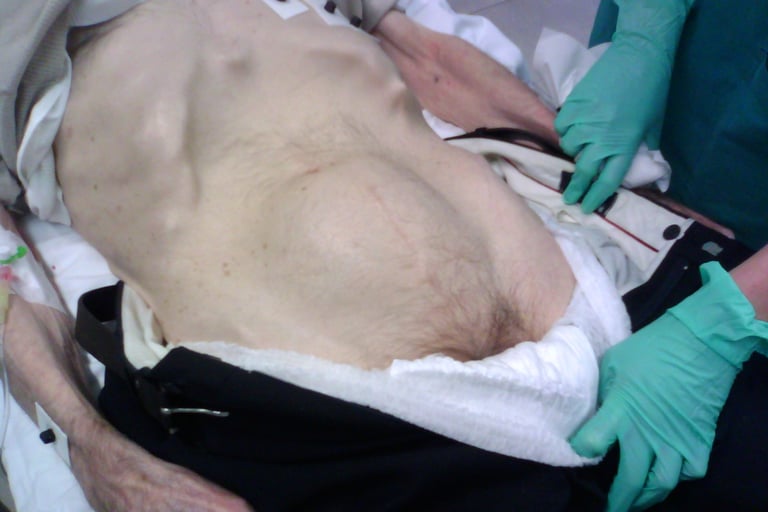
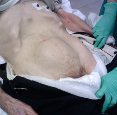
Image 1 - Distended bladder
Aetiology:
Mechanical - Constipation (via mechanical compression on the urethra), calculi in the bladder or urethra and foreign bodies
Men - BPH, Prostate cancer, urethral strictures, phimosis and paraphimosis
Women - prolapses, pelvic masses, gravid uterus
Infective - UTI
Medication-related - Anaesthesia, anticholinergics, Alpha agonists, antidepressants
Pain
Constipation
Neurological causes - Cauda equina
Examination:
Distended, palpable bladder with suprapubic tenderness.
DRE - Assess the prostate size and feel for any irregularities, as well as any faecal impaction causing retention. Assess anal tone and perianal sensation.
PV exam - Any prolapses or palpable pelvic masses.
Lower limb neurological examination, if indicated.
Management:
Routine blood tests (there is no indication for PSA in this context, as it will be raised secondary to retention).
Catheterise with a long-term catheter.
A 14 or 16Fr catheter in men
A 12 or 14Fr catheter in women.
Send a urine sample for dipstick and a M/C/S
Monitor the urine output and watch for diuresis if the residual urine volume post-catheterisation is >1000ml (diuresis is defined as a urine output of >200ml for 2 or more consecutive hours).
In uncomplicated cases, a TWOC can be arranged at the local urology intervention centre with 72 hours of tamsulosin cover (3 doses). However, some centres prefer a delayed approach with a TWOC in a week, which is sensible.
Start treatment for manageable causes of retention, such as antibiotics if suspecting a UTI or tamsulosin and finasteride if BPH was the likely cause of retention.
In women, an ultrasound TATV is useful to exclude pelvic pathology
Pitfalls:
Nothing's draining - Incorrect catheter placement, anuric patients and false positive bladder scans can cause this.
Anuric patients - there is no urine in the bladder. In this case, the cause of anuria must be sought, and rehydration commenced. A review by the renal team is recommended.Decompression haematuria - a common phenomenon where urine turns red or dark brown as a result of decompression of the kidneys once back pressure is relieved. This is transient, and patients should be reassured.
Chronic retention often has volumes >800ml, and patients may surprisingly not have much discomfort with these volumes of urine.
It is important to differentiate high-pressure chronic retention (altered renal function with hydronephrosis) and low-pressure chronic retention (minimal derangement and normal kidneys on imaging), as the former warrants a long-term catheter until the cause is managed. A history of nocturnal enuresis is a red flag symptom for high-pressure retention.
If blood tests are deranged and you suspect high-pressure chronic retention, arrange for an ultrasound scan KUB, correct diuresis at the rate of 50% of the hourly urine output with 0.9% sodium chloride, get daily body weights, lying and standing blood pressures and daily blood tests monitoring renal function and electrolytes.
Images:
Image 1 - Used under the Creative Commons licence - Frivadossi, CC BY-SA 3.0 <https://creativecommons.org/licenses/by-sa/3.0>, via Wikimedia Commons
UroMate
contact@uromate.com
© 2025. All rights reserved.
Disclaimer: The content on UroMate is intended for educational use by medical professionals only. It does not constitute professional medical advice, clinical guidelines, or a substitute for supervision or training. UroMate is not an official authority and accepts no responsibility for clinical outcomes. This site reflects UK clinical practice and may not apply elsewhere. Users must consult official sources such as NICE, EAU, or NHS protocols. Medical knowledge evolves, and while we strive for accuracy, content may not reflect the latest guidance. Not intended for patients or the general public. By continuing to use this site, you acknowledge and accept these terms.