The acute scrotum
This section covers:
Testicular torsion
Epididymo-orchitis
Torted hydatids
Testicular haematoma
Testicular torsion
A true urological emergency requiring an emergency exploration and fixation.
Acute onset of testicular pain, severe enough to cause nausea and vomiting at times.
Caused by a twist in the spermatic cord, which denies the testis of its blood supply, resulting in ischaemia and pain.
Torsion occurs in boys of any age. It can occur in the first year of life but most commonly in those aged 12-18 years, with peak incidence between 13-16 years.
There were 3,304 episodes of testicular torsion in England in 2013/14, of which 2,501 were in children [1].
Assessment:
History - Acute onset of unilateral testicular pain. May have a history of spontaneous pain that settled a few days ago, indicating a spontaneous torsion and detorsion.
Any scrotal swelling? If so, when did the swelling start? If it preceded the pain by a few days, an infection is more likely.
A history of previous scrotal operations is invaluable, and an operative note is golden, as a fixed testis is unlikely to twist on itself.
History of trauma? While most torsions can occur without a predisposing activity, up to 8% of torsions occur post-injury [2].
Ask about signs and symptoms of STIs - urethral discharge, sexual partners, dysuria.
Inspect - look for any gross hemiscrotal swelling, skin changes such as erythema, is the testis elevated or high riding? Is the testis in its normal vertical lie? Or is it horizontal?
Palpation:
-Cremasteric reflex (assessed on coughing or stroking the inner thigh) and observe for a normal response - elevation of the stimulated testis.
-Prehn's test is usually negative - elevation of the scrotum will not relieve pain in torsion.
-Always assess the hernial orifices for a cough impulse.
-Feel the testis, epididymis and cord. The testis and cord will be extremely tender, and most times, a patient with torsion will not tolerate palpation. At times, swelling around the testis may be noted, which is reactive fluid.
Adjuncts:
Routine blood tests and inflammatory markers
Urine dipstick - test for UTI that could have led to epididymo-orchitis
Ultrasound scans - no role unless immediately available, and will not delay surgical exploration
At its core, testicular torsion is a clinical diagnosis, and these adjuncts should not act as a guide to diagnose or rule out a torsion. A decision to operate should be made based on clinical judgement and long before investigations return. Have a low threshold to explore a young patient with a good history of torsion.
Management:
Emergency scrotal exploration as a category 1 procedure (keep the patient NBM.)
Orchidopexy (fixation of the testis to the dartos)
Orchidectomy if non-viable testis - dusky or dark with no evidence of reperfusion in 15- 20 minutes of active measures (placing in warm saline soaked gauze, administration of supplemental oxygen to the externalised testis)
Exploration of the contralateral hemiscrotum if the affected side was torted and fixation. (2% of testicular torsions are bilateral [3])
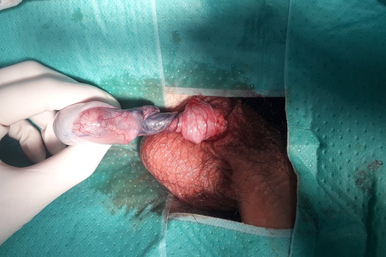
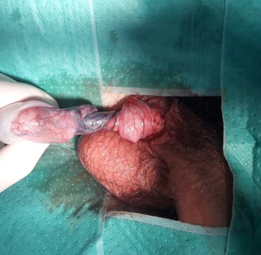
Image 1 - Torted spermatic cord
Torted testicular hydatid
A torted testicular appendage (hydatid of Morgagni) can present similarly to a true testicular torsion however, the following are key differences:
Tenderness at the upper pole of the testis.
A positive cremasteric reflex will be noted.
The hydatid may be palpated and, in some cases, may transilluminate through the scrotal skin as a ‘blue dot’.
Management of torted hydatids is conservative with rest, ice application and NSAIDs [4]. If in doubt, it is always a sensible idea to advocate for a scrotal exploration.
Epididymo-orchitis
Usually caused by the extension of infection from the urethra or bladder
Presents gradually as worsening pain and swelling. A history for STIs should be sought, and the patient should be encouraged to test for STIs with information on the nearest GUM clinic.
Aetiology:
In men <35 years of age, STIs (Chlamydia and gonorrhoea) are the most likely cause. A stat dose of Amoxicillin followed by doxycycline for 14 days
UTIs - E coli and Klebsiella
Amiodarone
Mumps - Check for immunisation history or previous infection
Behcet’s disease
Extrapulmonary TB
Unprotected anal intercourse - coliform organisms
Recent instrumentation/catheterisation
Assessment:
Swelling of the affected hemiscrotum.
Tenderness and a possibly bulky epididymis - palpate the tail, body and head.
Testicular tenderness is not uncommon.
Ask the patient to squeeze the penis and observe for urethral discharge.
See the examination under the testicular torsion section for more details and signs.
Investigations:
Urine dipstick is often times positive.
Urine for M/C/S.
Urethral swabs if urethral discharge noted - Gonorrhoea and chlamydia testing.
Refer to your local GUM clinic if the above cannot be arranged in hospital. The patient will also need counselling and contact tracing.
Blood tests - elevated white cells and inflammatory markers.
Management:
If an STI is suspected, IM Ceftriaxone (stat) + Doxycycline for 2 weeks.
If an STI is not suspected, 2 weeks of Ofloxacin or ciprofloxacin.
Advise the patient to avoid unprotected sex for 2 weeks after he and his partner (if applicable) complete the course of antibiotics to minimise the risk of re-infection.
Prescribe simple analgesia - codeine with paracetamol or ibuprofen.
Please consult your local antibiotic policy.
Advise patients to reattend the hospital or clinic if there is no improvement within 3 days or if symptoms worsen. At this point, an alternative diagnosis may have to be considered.
Testicular haematoma
Presents as a painful testis in a patient who has had trauma to the testis.
A history of trauma with acutely worsening swelling and pain is the most common setting.
Assessment:
Tender and swollen testis
A haematoma may be palpable if the testis has not ruptured
If the testis has ruptured, a haematocoele will be appreciated and the underlying testis will be difficult to palpate.
Erythema or early features of bruising of the scrotum may be noted
Investigation:
An ultrasound of the scrotum will help in determining whether there is testicular rupture - this will be noted as an irregular margin of the testis which corresponds to the ruptured tunica albuginea and the herniating testicular tissue
Management:
Conservative management can be trialled in small haematomas, pain that is controlled and in the absence of ruptured tunica albuginea. This includes a repeat ultrasound scan in 2 days.
Scrotal exploration and primary repair of the ruptured testis
A drain may be placed if a large haematocoele is noted
If the testis cannot be salvaged, an orchidectomy is warranted
Video on scrotal examination
Video 1 - Demonstration of scrotal examination
Other useful links:
https://patient.info/doctor/torsion-of-the-testis-pro
https://patient.info/doctor/epididymo-orchitis-pro
BAUS consensus on the management of testicular trauma - https://www.baus.org.uk/_userfiles/pages/files/professionals/sections/andrology/Testictrauma.pdf
Images:
Image 1 - Used under the Creative Commons licence - Javier.montero.arredondo, CC BY-SA 4.0 <https://creativecommons.org/licenses/by-sa/4.0>, via Wikimedia Commons
Video:
Video 1 - courtesy of Geeky medics. https://www.youtube.com/watch?v=KzOEc7X6RQw
References:
[1] https://eoeneonatalpccsicnetwork.nhs.uk/wp-content/uploads/2023/08/Children-Presenting-with-Acute-Scrotum.pdf
[2] Seng, Y.J. and Moissinac, K. (2000). Trauma induced testicular torsion: a reminder for the unwary. Journal of accident & emergency medicine, [online] 17(5), pp.381–2. doi:https://doi.org/10.1136/emj.17.5.381.
[3] Bokhari, A., Aldarwish, H., Alharbi, T., Alrashidi, Y., Alharbi, A. and Alsulami, L. (n.d.). Bilateral Testicular Torsion: A Systematic Review of Case Reports. Cureus, [online] 15(5), p.e38861. doi:https://doi.org/10.7759/cureus.38861.
[4] Pomajzl, A.J. and Leslie, S.W. (2020). Appendix Testes Torsion. [online] PubMed. Available at: https://www.ncbi.nlm.nih.gov/books/NBK546994/.
UroMate
contact@uromate.com
© 2025. All rights reserved.
Disclaimer: The content on UroMate is intended for educational use by medical professionals only. It does not constitute professional medical advice, clinical guidelines, or a substitute for supervision or training. UroMate is not an official authority and accepts no responsibility for clinical outcomes. This site reflects UK clinical practice and may not apply elsewhere. Users must consult official sources such as NICE, EAU, or NHS protocols. Medical knowledge evolves, and while we strive for accuracy, content may not reflect the latest guidance. Not intended for patients or the general public. By continuing to use this site, you acknowledge and accept these terms.